Patients
I'm told I need...

Leaflets are in Adobe Acrobat™ (PDF) format and can be downloaded, printed or saved on your computer. Most computers can read these files but, if yours cannot, you will need to download the free Acrobat Reader DC™ software by clicking the logo (left).
Insertion of a ureteric stent
NOTE: Some of the information provided contains graphic, medical images which individuals may find upsetting
Click here to use our feedback form & send us your comments about this section of the BAUS website; this will help us to improve it for the benefit of our patients.
How is the procedure performed?
This is normally performed under a brief general anaesthetic via a telescope passed into the bladder. A guidewire is inserted into the ureteric opening in the bladder and, using X-ray screening, is passed up into the kidney.
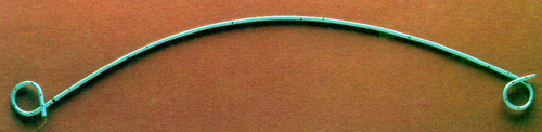
A ureteric stent (pictured above) is then pushed into the correct position and the guidewire removed once positioning has been confirmed by X-ray.
Video - Ureteric stent insertion
Features of this video (courtesy of Mr Nigel Bullock)
- Guidewire (pale green) inserted into the ureteric opening
- Stent (pale blue) pushed over the guidewire
- Hollow pusher (white) used to push the stent over the guidewire into the correct position
- Guidewire removed under X-ray control, leaving the stent correctly positioned