
Please note: This page is intended for healthcare professionals only. It is designed as a general educational guide and does not replace local guidance, senior clinical advice, or individual clinical judgement. Patients should not use this page as medical advice and should seek advice from an appropriate healthcare professional.

Male Lower Urinary Tract Symptoms (LUTS)

Male Lower Urinary Tract Symptoms (LUTS)

Lower urinary tract symptoms (LUTS) are among the commonest reasons for urological referral. Whilst many men presenting with LUTS are ultimately found to have benign prostatic enlargement (BPE), LUTS are symptoms rather than a diagnosis and may arise from multiple causes not only relating purely to urinary tract.

Your role is to recognise red flag symptoms and emergencies, identify the likely underlying cause, and arrange appropriate investigation or referral.

Classification of LUTS

LUTS are traditionally divided into storage, voiding and post-micturition symptoms. Patients commonly present with symptoms from more than one LUTS group and may not fit neatly into a single category.

1
Storage Symptoms – associated with difficulty storing urine. This can be related to bladder overactivity, polyuria or intrinsic bladder pathology (eg. stone, tumour):
  • Frequency
  • Urgency +/- urge incontinence
  • Nocturia
2
Voiding Symptoms – associated with difficulty passing urine. This is usually related to bladder outflow obstruction (BOO) or underactive bladder:
  • Weak stream
  • Hesitancy
  • Intermittency
  • Straining
  • Prolonged voiding
3
Post-Micturition Symptoms
  • Feeling of incomplete bladder emptying
  • Post-micturition dribbling
 

Causes of LUTS

LUTS may be caused by both urological and non-urological conditions. Think beyond the prostate.

Possible urological causes - these are well summarised by the EAU guidelines on “Non-neurogenic Male LUTS” 1 as seen in the graphic on the right.

Non-urological causes - diabetes, neurological disease (e.g. Parkinson's disease, stroke or dementia), heart failure, obstructive sleep apnoea and medication side effects.


Is this an emergency?

Acute LUTS

new or rapidly worsening symptoms should prompt consideration of:

  • Urinary tract infection
  • Acute prostatitis
  • Acute urinary retention
  • Distal ureteric stone
  • Neurological pathology (e.g. cauda equina syndrome or metastatic spinal cord compression)

Chronic LUTS

gradually progressive symptoms developing over months or years.

  • Usually, can be investigated electively.
  • You can start treatment if there is probable identified cause (for example, start Tamsulosin for BPE).
 
Red Flags
  • Visible haematuria
  • Hydronephrosis
  • Renal impairment
  • Nocturnal enuresis s. bedwetting (think HPCR)
  • New neurological symptoms
  • Abnormal DRE
  • Elevated PSA suspicious for malignancy
  • Unexplained weight loss
  • Recurrent UTI’s
 

Assessment

Immediate assessment during on-call:
1
History
  • Type of LUTS
    • Is it storage, voiding (obstructive) or mixed?
    • Spraying of urine can be indicative of a urethral stricture
    • Is there incontinence?
  • Onset
  • Red flag symptoms
  • Relevant medications
  • Past urological treatments / surgeries / relevant trauma / pelvic XRT
  • Neurological symptoms
  • Illicit drug use – especially Ketamine (read BAUS consensus on Ketamine bladder - HERE)
  • Other systems symptoms – Bowel issues? Heart failure? Diabetes? Sleep apnea?
2
Examination
  • Abdomen - palpable bladder?
  • Genital exam – phimosis? meatal stenosis? genital tumour?
  • DRE
    • Size – this is a rough estimate only and it can be difficult to determine size
      • Small / normal ~ 20-30cc
      • Severe >80cc
    • Nodules or cragginess – cancer?
    • Painful or fluctuant – prostatitis or abscess?
3
Investigations
  • Urine dip +/- MSU
  • Post-void residual (PVR)
    • Normal bladder capacity ~300-500ml
  • Bloods
    • U&E’s
    • WBC and CRP – if you think infection
    • PSA – IF APPROPRIATE
      • NICE guidelines on PSA testing – HERE
      • Patient leaflet on PSA – HERE
  • Imaging
    • USS or CT – should be done if this would change management or aid diagnosis:
      • Red flag symptoms
      • AKI
      • Unwell patient
      • Concerns about ureteric stone
    • MRI spine – if concern about new neurological symptoms and LUTS
Assessments that can be done in clinic:
1
Patient questionnaires
  • IPSS - a validated questionnaire used to quantify the severity of LUTS and monitor response to treatment. Available - HERE.
  • Bladder diary / frequency-volume chart
2
Uroflowmetry (flowrate) assessment
  • Provides an objective assessment of urinary flow
  • A reduced maximum flow rate (Qmax) may suggest:
    • Bladder outlet obstruction
    • Urethral stricture disease
    • Poor detrusor contractility
  • A low flow rate indicates a voiding problem but does not identify the cause and should be interpreted alongside symptoms, PVR and clinical findings
3
TRUS prostate size evaluation
4
Urinary tract imaging – NICE currently do not recommend imaging of the upper urinary tract for the routine assessment of uncomplicated LUTS. Many however, will favour this prior to managing the causes of LUTS
5
Flexible cystoscopy – useful to assess anatomy, exclude cancer and bladder stones, but not recommended for uncomplicated LUTS during initial work-up
6
Urodynamics – helpful if there is diagnostic uncertainty
 

Management

Management should target the underlying cause rather than the symptom alone.

Common Examples
Likely Cause Initial Management
BPE Lifestyle advice ± alpha-blocker
Urinary retention Catheterisation and TWOC planning
UTI Antibiotics according to local guidance
Acute prostatitis Antibiotics and supportive management
Urethral stricture Urology referral
Overactive bladder Bladder diary, lifestyle modification, bladder retraining and OAB treatments
Neurological pathology Urgent assessment (especially if acute)
  • Further information on individual conditions can be found in the relevant website sections.
  • BAUS patient information leaflet on “Male LUTS” - HERE
When to Refer

Consider referral to urology if there is:

  • Red flag symptoms
  • Recurrent urinary retention
  • Elevated PVR
  • Suspected urethral stricture disease
  • Failure of conservative or medical management
Take home message

Similar LUTS may arise from very different pathologies. A man with urgency and nocturia may have overactive bladder, diabetes, heart failure or sleep apnoea, whilst a man with a weak stream may have BPE, a urethral stricture or an underactive bladder.


Benign Prostatic Enlargement

Overview

Benign prostatic hyperplasia (BPH) is a histological diagnosis describing benign overgrowth of prostate tissue. In clinical practice, it is more useful to think in terms of:

Term Meaning
BPH Histological benign prostatic hyperplasia
BPE Benign prostatic enlargement
BPO Benign prostatic obstruction causing bladder outlet obstruction
  • BPE is common with increasing age, but not all men with an enlarged prostate have symptoms. Similarly, not all male LUTS are caused by the prostate.
  • The key question is whether the prostate is causing clinically significant obstruction or complications.
  • Prostate size, symptom severity and flow rate do not always correlate: a large prostate may cause few symptoms, while a smaller gland can still cause significant obstruction.
 

Epidemiology

Prostate reaches adult size (~ 20 +/- 6 g) between the ages of 21 and 30 years and generally remains stable unless BPH develops.

Prevalence of histological BPH:

  • 40s: ~8%
  • 51–60 years: ~50%
  • Large prostates are uncommon: >100 g prostate: ~4% of men aged >70 years

BPE develops predominantly within the transition zone, which surrounds the urethra and may lead to bladder outlet obstruction. In contrast, prostate cancer most commonly arises within the peripheral zone.


Assessment

Patients with suspected BPE/BPO should undergo a standard LUTS assessment as described in the LUTS section.


Management

1
Conservative – may be appropriate for mild or non-bothersome symptoms.

Advice includes:

  • Reduce evening fluid intake if nocturia is problematic
  • Reduce caffeine, alcohol and fizzy drinks if they worsen overactivity symptoms
  • Review contributing medications
  • Treat constipation
  • Double voiding if incomplete emptying is a problem
  • Weight loss if overweight

BAUS patient leaflet on male LUTS - HERE

2
Medical Management

Alpha-blockers (Tamsulosin, Alfuzosin, Doxazosin)

  • Mechanism: relaxes smooth muscles at the prostate and bladder neck, reducing dynamic obstruction and improving flow
  • DOES NOT reduce the size of prostate or stop the natural growth
  • They work relatively quickly, often within days to weeks
  • Common side effects:
    • Dizziness, postural hypotension, retrograde ejaculation

5-alpha reductase inhibitors (Finasteride, Dutasteride)

  • Mechanism: reduce conversion of testosterone to DHT and can reduce prostate volume over time
  • Symptom improvement is slow and may take 4–6 months
  • Prostate size can reduce by ~25%
  • PSA typically falls by ~ 50% after 6 months
  • Side effects:
    • Reduced libido, depression, erectile dysfunction, ejaculatory dysfunction, gynaecomastia or tender breasts, hot flushes

PDE5 inhibitors (Tadalafil 5mg OD)

  • Can be used for men with LUTS, particularly where erectile dysfunction coexists.
  • Does not reduce prostate size
  • Avoid concomitant nitrate therapy

Storage symptoms (solifenacin, trospium, mirabegron)

  • If storage symptoms predominate, consider whether the patient may have overactive bladder rather than pure obstruction. Antimuscarinics or beta-3 agonists may be used in selected patients, but caution is required if there is a high PVR or risk of retention.
3
Surgery – surgical treatment aims to relieve bladder outlet obstruction rather than simply reduce prostate size.

The SHO does not need to know the technical detail of every BPE procedure, but should understand when surgery may be considered.

See this section for common BPE procedures - HERE

Surgical treatment may be considered for:

  • Failed conservative or medical therapy
  • Recurrent acute urinary retention
  • Chronic low or high pressure urinary retention
  • Recurrent UTIs thought to be secondary to obstruction
  • Bladder stones
  • Recurrent haematuria attributed to BPE after appropriate investigation
  • Prior to radiotherapy for prostate cancer in men with significant LUTS or chronic retention (channel TURP)
Take home message
  • BPH, BPE and BPO are not interchangeable terms.
  • BPE is common with ageing, but not all enlarged prostates cause symptoms.
  • Not all male LUTS are caused by BPE/BPO.
  • Alpha-blockers improve dynamic obstruction but do not shrink the prostate.
  • 5-alpha reductase inhibitors reduce prostate volume but take months to work and lower PSA.
  • Surgery is considered for bothersome refractory symptoms or complications of obstruction.
 

Urethral Stricture Disease

Overview

A urethral stricture is a narrowing of the urethral lumen caused by scar tissue formation. This may result in progressive bladder outlet obstruction and subsequent recurrent infection, urinary retention and, in severe cases, upper tract deterioration.

Urethral strictures affect approximately 0.6% of men and are uncommon in women. Although they may occur at any age, they are more commonly diagnosed in older men.

Most strictures occur within the bulbar urethra.


Stricture Location

Urethral strictures are broadly divided into anterior and posterior strictures.

The majority of strictures occur within the anterior urethra. Stricture location is important because it influences both symptoms and treatment options.

Male urethra diagram showing prostatic, membranous, bulbar, penile, fossa navicularis and external urethral meatus segments
Anterior strictures

involve:

  • Meatal or submeatal (fossa navicularis)
  • Penile urethra
  • Bulbar urethra

Common causes include:

  • Idiopathic
  • Instrumentation (prior urological surgery, catheterisation, sounding etc.)
  • Balanitis xerotica obliterans (BXO)
  • Infection (particularly after STI)
  • Trauma (straddle injury, penile trauma)
Posterior strictures

involve:

  • Membranous urethra
  • Prostatic urethra
  • Bladder neck
    • Bladder neck contracture is not technically a urethral stricture but is commonly considered alongside posterior stricture disease

Common causes include:

  • Pelvic fracture urethral injury (PFUI)
  • Prostate surgery
  • Pelvic radiotherapy
 

When Should I Suspect a Stricture?

  • Gradually worsening weak stream
  • Spraying or split urinary stream
  • Obstructive LUTS or bladder outflow obstruction with a small prostate
  • Previous catheterisation or instrumentation
  • Previous pelvic trauma
  • BXO (could have history of circumcision)
  • Younger men presenting with LUTS, especially if there is a history of previous hypospadias surgery or points mentioned above
 
On-Call Relevance

The immediate priority is management of the acute problem. Definitive stricture treatment is usually arranged electively.

Common stricture-related presentations include:

  • Acute urinary retention
  • Difficult catheterisation
  • Recurrent urinary tract infection
 

Assessment

Patients with suspected urethral stricture disease should undergo a standard LUTS assessment as described in the LUTS section.

Particular attention should be paid to:

Retrograde urethrogram showing bulbar urethral stricture with bladder, stricture and tip of urethra labelled

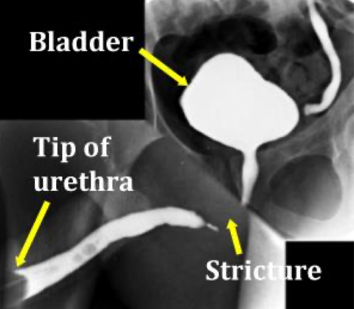
RUG of bulbar stricture

Retrograde urethrogram showing posterior urethral disruption with gap between urethra and bladder

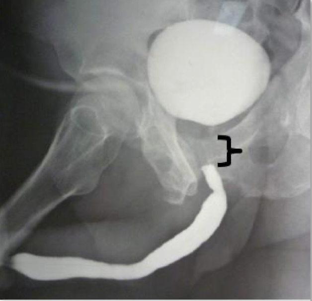
RUG of posterior urethral disruption

Cystoscopic view of urethral stricture with pinhole lumen during surgery, with stricture and knife blade labelled

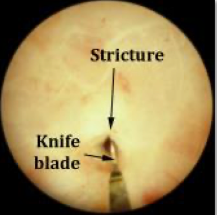
Cystoscopic view of stricture with pinhole lumen during surgery

1
History of
  • Previous catheterisation and endoscopic surgery
  • Pelvic trauma
  • Previous radiotherapy
  • Recurrent UTI’s
  • Spraying stream
  • Previous stricture treatment
  • Symptoms suggestive of BXO
2
During examination
  • External genitalia assessment
  • Examine perineum – look for scars and perineal abnormalities (eg. fistula)
3
Investigations

Investigations for strictures are usually done electively in outpatient setting. These include:

  • Urinalysis +/- MSU
  • Uroflowmetry and post-void residual (PVR)
    • May identify poor bladder emptying and chronic retention.
  • Renal Function
    • Check if retention or upper tract obstruction is suspected.
  • Upper Tract Imaging
    • Consider renal tract ultrasound in patients with:
      • Chronic retention
      • Elevated creatinine
      • Recurrent infections
  • Flexible Cystoscopy
    • Allows direct visualisation of the urethra and may identify:
      • Stricture location
      • Severity
      • Associated pathology
  • Retrograde Urethrogram (RUG)
    • Often considered the gold standard imaging investigation for urethral stricture disease.
    • RUG is particularly useful when planning surgery and may define:
    • It may also be used in the assessment of urethral trauma.
 
 
Difficult Catheterisation

Urethral strictures are a common cause of difficult catheterisation. If patient is known to have a stricture or you suspect it, then the principles are:

  • Never force a catheter!
  • Use a small diameter catheter (eg. 12Fr)
  • Use ample lubrication
  • Seek senior help EARLY if resistance is encountered
  • Repeated traumatic attempts may worsen the stricture and create false passages

Further information is available in the Difficult Catheterisation section.


Management Principles

Management depends on:

  • Stricture length
  • Stricture location
  • Previous treatments
  • Patient factors

Short, uncomplicated strictures may be suitable for endoscopic treatment, whereas recurrent or complex strictures often require reconstructive surgery.

See further information on procedures - HERE

A. Endoscopic Treatments
Urethral dilatation
Optical urethrotomy (DVIU)
Optilume drug-coated balloon

These may be appropriate for selected short strictures but recurrence remains common.

B. Reconstruction

This is usually done for patient with recurrent strictures suitable for surgery

Posterior strictures

These cases are often more complex and are typically managed in specialist reconstructive centres.

Anterior strictures
  1. Meatoplasty
  2. Urethroplasty
    • Considered the gold standard curative treatment for recurrent or complex strictures
    • This involves open reconstruction of the urethra and may utilise local tissue or buccal mucosal grafts.
C. Urinary diversion

In cases if restoration of normal urethral voiding may not be possible or desirable.

Long term urinary catheter (urethral or SPC)
Perineal Urethrostomy
Formation of ileal conduit
D. Stricture control
Intermittent self-dilatation (ISD)

For patients who prefer to avoid major surgery or are not suitable for definitive reconstruction they can manage their stricture with regular self-dilatation.

When to Refer?

Refer to urology if there is:

  • Suspected urethral stricture disease
  • BXO involving the urethral meatus
  • Difficult catheterisation
  • Persistent LUTS despite treatment
 

References

  1. European Association of Urology. EAU Guidelines on Non-neurogenic Male LUTS (2026)EAU LUTS Guidelines
  2. European Association of Urology. EAU Guidelines on Urethral StricturesEAU Urethral Stricture Guidelines
  3. NICE Guideline CG97. Lower Urinary Tract Symptoms in Men: ManagementNICE LUTS Guideline (CG97)
  4. NICE Clinical Knowledge Summary. Prostate Cancer – AssessmentNICE CKS Prostate Cancer Assessment
  5. Berry SJ, Coffey DS, Walsh PC, Ewing LL. The Development of Human Benign Prostatic Hyperplasia with Age. J Urol. 1984;132:474–479.Berry et al. 1984 (PubMed)
  6. Bugeja S, Payne SR, Eardley I, Mundy AR, on behalf of BAGURS. The Standard for the Management of Male Urethral Strictures in the UK: A Consensus Document. DOI: 10.1177/2051415820933504
  7. BAUS/BAGURS Urethral Stricture Consensus Document
  8. Belal M, et al. British Association of Urological Surgeons Consensus Statements on the Management of Ketamine Bladder Syndrome. BJU International. 2024. DOI:10.1111/bju.16404
  9. BAUS Consensus Statement on Ketamine Bladder Syndrome
  10. BAUS Patient Information Leaflets