 You will see a QR code graphic (right) which, when scanned, will also take you directly to the relevant document on this website.
Below the graphic there is a short URL which can be typed into a web browser, and will take you to the same document (if no QR code scanner is available).
This graphic also appears on the print versions of the relevant patient information leaflet, from where it can also be scanned.
The QR code graphic can be deployed to:
- patient (or clinician) correspondence;
- posters in your department; and
- your own information leaflets.
(i.e. anywhere where the recipient may wish to download the document to a mobile device and read it off-line).
To save the QR code for your own use, right click the graphic, then select the option to save/download it to your computer
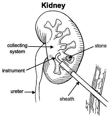
How is the kidney reached?
Under general anaesthetic, a telescope is us
ed to insert a small ureteric catheter from the bladder up to the kidney. This allows dye to be injected into the kidney and prevents stone fragments from falling down the ureter towards the bladder to cause a blockage.
The kidney is then punctured with a needle, using X-ray or ultrasound screening, so that a guidewire can be inserted into the drainage system of the kidney. This guidewire is used to "stretch up" an opening into the kidney, approximately 1cm wide, through which instruments can be inserted to inspect the inside of the kidney (as in the diagram, righ).
How is the stone broken & removed?
Small stones (less than 1cm across) can often be removed intact.
Larger stones need to be broken up using a laser (pictured right), an ultrasonic probe or a vibrating metal probe which breaks the stone like a pneumatic drill.
The larger stone fragments are picked out with forceps and the smaller fragments either wash out during the procedure or are sucked out using the ultrasound probe.
Click here to view an endoscopic video of ultrasound stone fragmentation, courtesy of Mr Oliver Wiseman