 You will see a QR code graphic (right) which, when scanned, will also take you directly to the relevant document on this website.
Below the graphic there is a short URL which can be typed into a web browser, and will take you to the same document (if no QR code scanner is available).
This graphic also appears on the print versions of the relevant patient information leaflet, from where it can also be scanned.
The QR code graphic can be deployed to:
- patient (or clinician) correspondence;
- posters in your department; and
- your own information leaflets.
(i.e. anywhere where the recipient may wish to download the document to a mobile device and read it off-line).
To save the QR code for your own use, right click the graphic, then select the option to save/download it to your computer
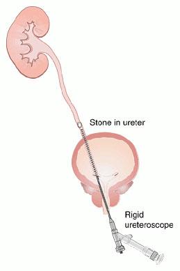
How is the procedure performed?
This is normally performed under a general or spinal anaesthetic.
The bladder is examined first using a small telescope. A guidewire is inserted into the opening of the ureter and passed up into the kidney; correct positioning is confirmed by an "on-table" X-ray.
A thin rigid instrument (see diagram, right) or a flexible telescope is then inserted and the guidewire is followed to allow inspection of the whole ureter and, if necessary, the kidney itself.
Any abnormality can be biopsied or removed whilst stones can be extracted or fragmented with a laser. A ureteric stent may be inserted at the end of the procedure, depending on what procedure has been performed.
Click here to view an endoscopic video of the procedure, showing polyps and swelling due to stone disease with infection: video courtesy of Mr Nigel Bullock